


Digestion and Absorption
Digestion : The process in alimentary canal by which the complex food is converted mechanically and biochemically into simple substances suitable for absorption and assimilation. Food : A substance which on taken and digested in the body provides mate- rials for growth, repair, energy, reproduction, resistance from disease or regulation of body processes.
DIGESTIVE SYSTEM :
Includes:
- Alimentary canal
- Digestive glands or associated glands.
Alimentary canal :
- The alimentary canal begins with mouth and ends with anus.
- Mouth leads to buccal cavity or oral cavity.
- Oral cavity has teeth and muscular tongue.
- Each tooth embedded in a socket of jaw bone: such attachment called thecodont.
- Diphyodont : human has two sets of teeth in their life time:
- Milk teeth or deciduous teeth
- Permanent teeth.
- Heterodont : teeth are of unequal shape and size.
- Incisor (I)
- Canine (C)
- Premolar (PM)
- Molar (M).
- Dental formula : arrangement of teeth in each half of the upper jaw and lower jaw.
- Dental formula of human adult is

- The hard chewing surface of the teeth made up of enamel.
- The tongue is a freely movable muscular organ attached to the floor of the oral cavity by the frenulum.
- The upper surface of tongue has small projections called papillae, some of which bears taste buds.
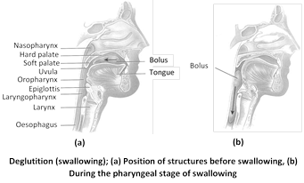
- The oral cavity leads into a short pharynx which serves as a common passage for food and air.
- Oesophagus and the trachea open into the pharynx.
- Opening of wind pipe or trachea called glottis, and that of oesophagus is called gullet.
- The cartilaginous epiglottis prevents the entry of food into the glottis during swallowing.
- Oesophagus connects pharynx with stomach.
- Opening of oesophagus is regulated by gastro-oesophageal sphincter.
- The stomach has three parts:
- Cardiac: into which oesophagus opens.
- Fundus: air filled portion of stomach.
- Pyloric: portion opens into the small intestine.
- Small intestine distinguished into three parts:
- Duodenum: ‘U’ shaped first part.
- Jejunum: longer, coiled middle portion.
- Ileum: highly coiled posterior part.
- The opening of stomach into the duodenum is guarded by pyloric sphincter.
- Large intestine consists of three parts:
- Caecum
- Colon
- Rectum.
- Caecum is a small blind sac which hosts some symbiotic micro-organisms.
- Caecum has a finger-like blind tubular projection called vermiform appendix.
- The Caecum opens into colon, which has three distinct part-
- Ascending colon
- Transverse colon
- Descending colon
- The descending colon opens into rectum which opens to out through anus.
Histolology of alimentary canal :
- Alimentary canal from oesophagus to rectum has four layers.
- Serosa.
- Muscularis.
- Sub mucosa.
- Mucosa.
- Serosa is the outermost layer and is made up of a thin mesothelium with some connective tissues.
- Muscularis is formed by smooth muscles arranged outer longitudinal and inner circular layers.
- Sub-mucosa is formed by loose connective tissues containing nerves, blood and lymph vessels.
- Mucosa is the innermost layer made of endothelium.
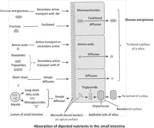
- Mucosa forms irregular folds (rugae) in the stomach and small finger like folding called villi in the small intestine.
- The cells lining the villi produce numerous microscopic projections called microvilli giving a brush border appearance.
- These modifications increase the surface area for absorption.
- Villi are supplied with a network of capillaries and a central lymphatic vessel called lacteal.
- Epithelial cells of mucosa contain secretory cells which secretes digestive enzymes.
- Mucosa also forms glands in the stomach (gastric gland)
- Mucosa forms crypts in between the bases of villi in the intestine called Crypts of Lieberkuhn.
*crypts of Lieberkühn (intestinal glands) Tubular glands that lie between the finger-like projections (see villus) of the inner surface of the small intestine.
The process of digestion involves following steps –
(1) Ingestion : It is the intake of food most of the animals capture the prey/food with the help of mouth or tongue.
(2) Mastication : The process occurs in the buccopharyngeal cavity of mammals with the help of teeth. During this process food is broken down into small pieces, which increases its surface area. In frog teeth are not meant for mastication but prevents the escape of prey from mouth.
(3) Deglutition / swallowing : The passage of food from buccopharyngeal cavity to oesophagus/stomach. In mammals bolus of the masticated food is formed in buccopharyngeal cavity which easily slides into oesophagus. It is a voluntary reflex mechanism. Peristalsis is alternative contraction and relaxation of circular and longitudinal muscles produces the wave of contraction due to which the food passes from front to backward direction in the lumen of alimentary canal. The phenomenon is called as peristalsis. Beside alimentary canal, it is also found in vas deference, ureter etc. Peristalsis is maximum in oesophagus and minimum in rectum.
Antiperistalsis is the peristaltic wave occurs in the reverse direction. It occurs in alimentary canal and results in vomiting. The phenomenon is called as “Regurgitation”.
(4) Digestion : The process by which complex food is converted into simple food with the help of digestive enzymes. The process of digestion in mammals starts in buccopharyngeal cavity.
(i) Digestion in buccopharyngeal cavity : In buccopharyngeal cavity of mammals only starch is digested which is 5% of total food or
(ii) Digestion in stomach : Chiefly proteins is digested in stomach.
(iii) Digestion in small intestine : All three component carbohydrates, proteins and fats digested in small intestine with the help of enzymes secreted by pancreas and intestinal glands.
(5) Absorption : Ingestion and digestion are the first two phases of the physiological processes occuring in the alimentary tract. The third phase is that of absorption by which the digested nutrients are absorb through the wall of gut into blood.
(i) Absorption from the mouth : Normally, there is no absorption from the mouth, but a few drugs may be absorbed into the blood through the mucous membrane, if allowed to dissolve under the tongue, e.g., isoprenaline, glyceryl trinitrate.
(ii) Absorption from the stomach : In the stomach, absorption takes place to a limited degree. The only substances normally absorbed from the stomach are some water, glucose and considerable amounts of alcohol. These substances are absorbed through the walls of the stomach into the venous circulation. Although iron absorption takes place in the small intestine, it is dissolved out of foods most effectively in the stomach in the presence of
(iii) Absorption from the small intestine : The small intestine is the main absorptive organ. About 90% of the ingested foodstuffs is absorbed in the course of passage through the small intestine.
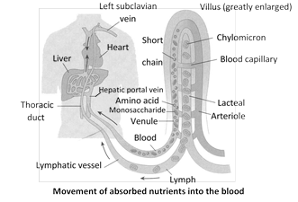
There are two general pathways for the transport of materials absorbed by the intestine; the veins of the hepatic portal system which lead directly to the liver; and the lymphatic vessels of the intestinal area, which eventually lead to the blood by way of the lymphatic system and the thoracic duct.
Absorption of carbohydrates : The products of carbohydrate digestion is absorbed from the intestine into blood of the portal venous system in the form of monosaccharides, chiefly the hexoses (glucose, fructose, mannose and galactose).
Absorption of amino acids and protein : It is probable that under normal circumstances the dietary proteins are almost completely digested to their constituent amino acids and that these end products of protein digestion are then actively transported from the intestine into the portal blood. Surplus amino acids are also withdrawn from portal blood by liver cells and deaminated into ammonia and keto acids. The ammonia is converted to urea and released into blood for excretion by kidneys, while the keto acids are converted to glucose or pyruvic acid and utilized for energy-production or for storage as glycogen and fat.
Absorption of fats : The dietary fat is digested, by the action of the pancreatic lipase present in the intestine, partially into glycerol and fatty acids and partially to split products such as monoacyl glycerols. These products of fat digestion enter the mucosal cells of the small intestine in the forms of micelles, fatty acids and glycerol.
By the lacteals, the fat is carried to the cisterna chyli (meaning 'the receiver of the chyle') and then by the thoracic (lymph) duct to the left branchiocephalic vein, where it enters the blood. The lymph reaching the thoracic duct from the intestines contains an excess of fat giving it a milky appearance. It is called chyle. In this way, fatty acids and glycerol are eventually brought into the blood stream and so, by a circuitous route, to the liver. In the liver, they are reorganized and recombined to form human fat.
Absorption of vitamins : Water-soluble vitamins like members of B complex (except
(iv) Absorption in large intestine : About 100-200 ml. of the water of undigested food is absorbed in the colon. It helps in maintaining the body water level. Some amount of mineral salts and vitamins are also absorbed. The symbiotic bacteria (E. coli) present in the large intestine, converts the inactive vitamins into active forms (i.e., they synthesizes vitamins (vitamin B complex and vitamin K) which are absorbed.
(6) Assimilation : Conversion of absorbed food into active cytoplasm within cell is called as assimilation.
(7) Faeces formation : The phenomenon occurs in colon due to absorption of water, salts, minerals and vitamins. The peristalsis in colon also helps in faeces formation.
(8) Egestion / defaecation : The elimination of faeces from the elementary canal is called egestion or defaecation. The faeces is waste matter discharged from the alimentary canal.
Pseudo-rumination or coprophagy : Animals swallows night faeces and recycle it through the gut to complete the digestion of cellulose and, making full use of their food. This habit is called coprophagy. Example – Rabbit.
Summary of physiology of digestion Major gastrointestinal enzyme in mammals
Name of gland
|
Name of digestive juice & optimum pH
|
Name of enzyme
|
Site of action
|
Substrates
|
Products
|
Salivary glands
|
Saliva (6.3 - 6.8)
|
Ptyalin / Salivary amylase
|
Mouth
|
Starch, dextrins, glycogen
|
Dextrins, maltose, isomaltose and limit dextrin.
|
Gastric glands
|
Gastric Juice (1-3)
|
Pepsin
|
Stomach
|
Proteins, casein (Milk)
|
Peptones, paracasein (curd).
Proteoses
|
Rennin
|
Stomach
|
Casein
|
Paracasein
| ||
Gastric lipase
|
Stomach
|
Fats
|
Fatty acid and Glycerol.
| ||
Liver
|
Bile juice (7.6-8.6)
|
No enzymes
|
Duodenum
|
Fat
|
Makes the food alkaline, emulsifies fat and kills the harmful bacteria.
|
Liver
|
Bile ( 7.6 - 8.6)
|
No enzyme but useful digestive juice, provides alkaline medium, stops the action of HCl. Emulsifies fats and kills harmful bacteria.
| |||
Pancreas
|
Pancreatic Juice (8.8)
|
Amylase/Diastase
|
Small intestine
|
Starch,
dextrins, glycogen.
|
'Limits' dextrins, maltose, isomaltose.
|
Trypsin
|
Small intestine
|
Proteins, Chymotry-psinogen (inactive) procarboxy pept- idases (inactive) Fibrinogen (blood) Casein (milk)
|
Peptides, Chymotrypsin (active) carboxy peptidases (active) Elastase (active), Fibrin (clot) Paracasein (curd)
| ||
Chymotrypsin
|
Small intestine
|
Peptones
|
Peptides
| ||
Carboxypeptidases
|
Small intestine
|
Peptides
|
Smaller peptides and Amino acids.
| ||
Lipase / Steapsin
|
Small intestine
|
Triglycerides
|
Mono-glycerides, fatty acids
| ||
DNAase
|
Small intestine
|
DNA
|
Deoxyribonucleotides
| ||
RNAase
|
Small intestine
|
RNA
|
Ribonucleotides
| ||
Intestinal glands
|
Intestinal Juice
(7.5-8.3)
|
Enteropeptidase (enterokinase)
|
Small Intestine
|
Trypsinogen (inactive)
|
Trypsin (active)
|
Aminopeptidase
|
Small Intestine
|
Peptides
|
Smaller peptides and amino acid
| ||
Dipeptidases
|
Small Intestine
|
Dipeptides 'Limit dextrins'
|
Amino acids
| ||
Isomaltase
|
Small Intestine
|
Isomaltose
|
Glucose
| ||
Maltase
|
Small Intestine
|
Maltose
|
Glucose
| ||
Sucrase/Invertase
|
Small Intestine
|
Sucrose
|
Glucose, fructose
| ||
Lactase
|
Small Intestine
|
Lactose
|
Glucose, galactose
| ||
Lipase
|
Small Intestine
|
Triglycerides
|
Monoglycerides, fatty acids
| ||
Nucleotidase
|
Small Intestine
|
Nucleotides
|
Nucleosides, inorganic phosphate
| ||
Nucleosidase Phosphorylases
|
Small Intestine
|
Nucleosides Phosphate
|
Purine, pyrimidine, pentose, phosphate
| ||
(9) Hormonal control of digestion : Activities of digestive tract are coordinated by nervous and endocrine systems. Sight and smell of food stimulates nervous system which induces the salivary glands to produce large quantity of saliva, stomach to release its hormone gastrin and intestine to produce intestinal hormones. Other hormones are produced in sequential order. All of them are polypeptide hormones.
Gastrointestinal hormones in mammals
Hormone
|
Source
|
Stimulus for secretion
|
Target organ
|
Action
|
Gastrin
|
Mucosa of pyloric stomach
|
Distension of stomach on food entry
|
Stomach
|
Stimulates secretion of gastric juice.
Constricts cardiac sphincter.
|
Enterogastrone
|
Duodenal epithelium
|
Chyme entry into duodenum
|
Stomach
|
Slows gastric contractions to delay its emptying.
Stops secretion of gastric juice.
|
Secretin
|
Duodenal epithelium
|
Acidic chyme entry into duodenum
|
Pancreas
Liver
Stomach
|
Release of sodium bicarbonate in pancreatic juice.
Steps up secretion of bile.
Inhibits secretion of gastrin.
|
Cholecystokinin
(Pancreozymin)
|
Duodenal epithelium
|
Presence of fats in duodenum
|
Pancreas
Gall Bladder
|
Release of enzymes in pancreatic juice.
Release of bile from gall bladder.
|
Villikinin
|
Intestinal epithelium
|
Food in small intestine
|
Intestine
|
Accelerates movements of villi.
|
Duocrinin
|
Intestinal epithelium (Duodenal mucosa)
|
Acidic chyme in intestine
|
Intestine (Brunner's gland)
|
Release of viscous mucous from Brunner's glands.
|
Enterocrinin
|
Intestinal epithelium
(Duodenal mucosa)
|
Acidic chyme in intestine
|
Intestine (crypts of Lieberkuhn's)
|
Release of enzymes from Lieberkuhn?s crypts.
|
Digestive glands :
- The digestive glands associated with the alimentary canal includes-
- Salivary gland
- Liver
- Pancreas.
- There are three pairs of salivary gland present in the buccal cavity.
- Parotid gland (below internal ear)
- Sub-maxillary / submandibular (below lower jaw)
- Sub-lingual (below tongue)
- All salivary glands produce saliva into the buccal cavity.
Liver :
- Largest gland of the body weighing about 1.2 to 1.5 kg in adult.
- Located below diaphragm and has two lobes.
- Structural and functional unit of liver is the hepatic lobules.
- Hepatic lobules consist of hepatic cells arranged in the form of cords.
- Each lobule is covered by a thin connective tissue sheath called Glisson’s capsule.
- The bile secreted by the hepatic cells passes through the hepatic ducts and stored in the gall bladder in concentrated form.
- Bile from the gall bladder is transported by cystic duct.
- Cystic duct along with hepatic duct forms the common bile duct.
- Bile duct joined with pancreatic duct to form hepato-pancreatic duct which open into the duodenum.
- Hepato-pancreatic has a swelling portion called ampulla of Vater; the opening is guarded by sphincter of Oddi.
Pancreas :
- Pancreas is a compound myxocrine gland (both exocrine and endocrine) elongated organ situated between the limbs of ‘U’ shaped duodenum.
- The exocrine aciner cells secrete pancreatic juice containing enzymes.
- The endocrine Islets of Langerhans secrete hormones like insulin and glucagon.
DIGESTION OF FOOD :
- Digestion is accomplished by mechanical and chemical process.
In the buccal cavity :
- Buccal cavity performs two major functions;
- Mastication of food.
- Facilitation of swallowing.
- The teeth and tongue with the help of saliva masticate and mix up the food.
- The saliva composed of ;
- Electrolytes (Na+, K+, Cl- HCO-3)
- Enzyme- salivary amylase or ptyalin.
- Lysozyme.
- About 30% of starch is hydrolyzed into disaccharide (maltose) by salivary amylase in optimum pH 6.8).
- Lysozyme acts as antibacterial agent preventing infections.
- Mucus in the saliva helps in lubricating and adhering the masticated food particle into a bolus.
- The bolus is then passed into oesophagus through pharynx by swallowing or deglutition.
- By peristalsis the bolus from the oesophagus passed into the stomach.
In the stomach :
- The mucosa of stomach has gastric glands.
- Gastric glands have three major types of cells namely –
- Mucus neck cells – secretes mucus.
- Peptic or chief or zymogen cells – secretes proenzymes pepsinogen.
- Parietal or oxyntic cells – secretes HCl and castles intrinsic factor (factor essential for absorption of vitamin B12)
- The stomach stores the food for 4-5 hours.
- The food mixed with the acidic gastric juice and form chyme.
- Pepsinogen converted into active pepsin in presence of HCl.
- Active pepsin converts proteins into proteose and peptones (peptides).
- Mucus and bicarbonate ions play important role in lubrication and protection of mucosal epithelium from excoriation by HCl and active enzymes.
- HCl provides the acidic pH of stomach (pH1.8)
- Rennin is an enzyme present in gastric juice helps in digestion of milk proteins.
- Small amount of lipases are present in gastric juice helps in digestion of fats.

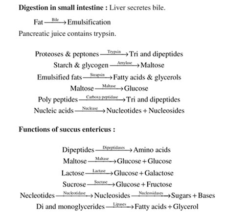
In the intestine :
- Important secretion added to the intestine during digestion:
- Bile juice.
- Pancreatic juice.
- Intestinal juice or succus entericus.
- The pancreatic juice contain following enzymes:
- Trypsinogen
- Chymotrypsinogen
- Procarboxypeptidase.
- Amylases
- Lipases
- Nucleases.
- Trypsinogen is activated by an enzyme, enterokinase secreted by intestinal mucosa into active trypsin.
- Active trypsin activates other enzymes in the pancreatic juice in the intestine.
- The bile released into the duodenum contains –
- Bile pigments (bilirubin and bili-verdin)
- Bile salts. (Bicarbonate, tourocholate, glycolate)
- Cholesterol and
- Phospholipids.
- Bile salt helps in emulsification of fat, i.e. breakdown fats into small micelles.
- Bile also activates lipases.
- The intestinal mucosa contains goblet cells which secrete mucus.
- The secretion of brush border cells of intestinal mucosa and the goblet cells constitute the intestinal juice or succus entericus.
- The intestinal juice contains variety of enzymes –
- Disaccharidases (maltase, lactase and invertase)
- Dipeptidases.
- Lipases.
- Nucleosidases.
- Sub-mucosal glands (Brunner’s glands) also secrete alkaline fluid to counter act acidic chyme before secretion of bile and pancreatic juice.
ABSORPTION OF DIGESTED PRODUCTS :
- Absorption is the process by which the end product of digestion passes through the intestinal mucosa into the blood or lymph.
- Absorption is carried out by passive, active or facilitated transport mechanism.
- Glucose, amino acids and electrolytes are absorbed by simple diffusion into the blood in the concentration gradient.
- Fructose and some amino acids absorbed with the help of carrier ions like Na+. This is called facilitated diffusion.
- Active transport of digested food and electrolytes takes place against the concentration gradients hence require energy.
Absorption of fatty acid and glycerol.
- Fatty acids and glycerol being insoluble cannot be absorbed into blood.
- They are transported into mucosal epithelium and triglycerides are formed.
- Triglycerides are covered by a protein coat to form small fat globules called chylomicron, which are incorporated into the lacteal in the villi.
- These lymphatic vessels ultimately release the absorbed substances into the blood stream later on.
Assimilation and egestion :
- The absorbed substances finally reach the tissues which utilize them for their activities. This process is called assimilation.
- The digestive wastes, solidified into coherent faeces in the rectum and removed to outside periodically by the process called defaecation.
Nutritional disorders : Every organism requires an adequate supply of nutrients in proper proportion in their diet for proper growth and development. There are two types of nutritional disorders
(1) Diseases due to over nutrition
(i) Fluorosis : Caused due to excess of fluorides. It results in tooth and bone decay.
(ii) Obesity : This is over-nutritional disorder. It is caused when 'energy inputs exceeds energy output'. It results in deposition of excess fat in the body.
(iii) Constipation : Slow movement of faeces down the large intestine causes accumulation of dry and hard stool is colon. It is generally caused by irregular bowel habits.
(iv) Diarrhoea : Rapid movement of faecal matter down the large intestine causes loose stools called diarrhoea. It may be also caused by viral or bacterial infections of intestinal tract, particularly of large intestine and by nervous tension.
(v) Piles or haemmorhoids : Enlargement of the anal veins. It may be either hereditary or may be caused due to rapid changes in the diet.
(vi) Hypercholesterolemia : Caused due to excess of saturated fats like butter, ghee, hydrogenated vegetable oils and eggs etc. It results in increased level of cholesterol in blood, arteriosclerosis, coronary thrombosis, heart attack etc.
(vii) Hypervitaminosis A : It results in loss of appetite, body hairs, painful swelling etc.
(viii) Hypervitaminosis D : It results in deposition of calcium ion in the soft tissues of the body.
Indigestion :
- The food is not properly digested leading to a feeling of fullness.
Causes are inadequate enzymes secretion, anxiety, food poisoning, over eating and spicy food.
(2) Diseases due to deficiency of nutrition (malnutrition)
Name of the Deficiency
|
Deficient Nutrient
|
Symptoms
|
Anaemia (microcytic)
|
Fe
|
Haemoglobin and number of erythrocytes gets reduced.
|
Megaloblastic anaemia
|
Folic acid and
|
Presence of immature erythrocytes in blood.
|
Pernicious anaemia
|
Vitamin
|
Immature RBC without Hb. This may be fatal unless treated with vitamin
|
Xerophthalmia
|
Vitamin A
|
Thickened, keratinised, opaque ulcerated cornea. Prime cause of blindness in India, especially among children.
|
Night Blindness
|
Vitamin A
|
Less rhodopsin in rod cells of retina. So no vision in dim light.
|
Rickets (in children)
|
Vitamin D
|
Weak, soft, thin bones due to poor deposition of Ca and P. Bent long bones and painful swelling on wrist, elbow and knee joints.
|
Osteomalacia (adults)
|
Vitamin D
|
Weak bones of vertebral column, pelvis gets bent and deformed by body weight.
|
Sprue
|
Folacin
|
Ulceration of mouth, inflammation of bowel, indigestion, diarrhoea, weakness.
|
Pigeons breast
|
Incomplete ossification at the end of limb bone, deformed ribs leading to pigeons breast.
| |
Beri ? beri
|
Vitamin
|
Reduces aerobic carbohydrate metabolism. So peripheral nerves inflammed causing pain, numbness and weakness of limb muscles. Paralysis.
Fluid accumulation in tissues or oedema of hands and legs. Cardiac oedema.
|
Scurvy
|
Vitamin C
|
Fragile blood vessels because of defective collagen fibres in their walls. Bleeding gums, teeth fall, bones fragile. Wound healing delayed, vitamin C recommended in serious injury.
|
Bleeding disease
(Hypoprothrombin anaemia)
|
Vitamin K
|
Delayed blood clotting (s) so profuse bleeding.
|
Marasmus
|
Protein / Malnutrition
|
Growth and replacement of tissue proteins imparted so emaciated body with their limbs and prominent ribs, dry, thin and wrinkled skin, Diarrhoea. It affects infants under one year of age.
|
Kwashiorkor
|
Protein
|
Wasting muscles, thin limbs, Retarded growth of body and brain, Oedema, Diarrhoea. It commonly affects babies between
|
Pellagra
|
Nicotinamide
|
Swollen lips, thick pigmented skin of hands and legs. Irritability.
|
Osteoporosis
|
Weakning of bones, tooth decay.
| |
Goitre
|
Enlargement of thyroid gland.
| |
Muscular cramps
|
Pulling of muscles due to dehydration.
| |
Dental cramps
|
Fluorine
|
Tooth decay.
|
0 Comments:
Post a Comment